

Full-body MRI: what it can detect and when the evidence says it helps
The essentials
- Full-body MRI has a role backed by evidence in specific high-risk groups, such as people with Li-Fraumeni syndrome (an inherited cancer predisposition) or suspected multiple myeloma.[1][2]
- In Li-Fraumeni syndrome, a pooled analysis of 578 people found a new, localized primary cancer in about 7% at the first scan.[1]
- In people with no symptoms or risk factors, no study has shown that scanning lowers mortality, and the American College of Radiology does not recommend screening in that group.[3][4]
- Because it images so much tissue at once, incidental findings are common: most are not cancer, but they can lead to more tests.[5]
What a full-body MRI is
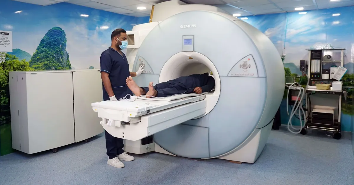
Magnetic resonance imaging builds pictures with magnetic fields and radio waves, not X-rays.[6] A full-body scan covers a large part of the body in one session, and the exam usually takes 60 to 90 minutes.[4] Some MRI exams use a contrast agent (gadolinium) given through a vein; whether yours needs one depends on the protocol.[6]
There is no single protocol: each center decides which regions and sequences to include, and that changes a lot about what gets found. To reduce this variation, an international panel proposed ONCO-RADS in 2021, a system that standardizes how a full-body MRI is acquired, interpreted and reported when it is used for cancer detection in higher-risk people.[7]
Where the evidence is strongest: people at high cancer risk
Li-Fraumeni syndrome
Li-Fraumeni syndrome is an inherited condition caused by pathogenic variants in the TP53 gene. People who have it face a very high lifetime risk of cancer, including sarcomas, breast cancer, brain tumors, leukemia and adrenocortical carcinoma, often at young ages.[8] It is the group with the strongest evidence, summarized in three sources:
- Prospective study (Villani, 2016). It followed 89 carriers of TP53 variants at three centers in Canada and the United States. The 59 who followed a surveillance protocol (whole-body MRI, brain and breast MRI, mammography, abdominal and pelvic ultrasound, colonoscopy and lab tests) were monitored for a median of 32 months, and 40 asymptomatic tumors were detected in 19 of them (32%). Five-year overall survival was 88.8% with surveillance and 59.6% without.[8]
- Meta-analysis (Ballinger, 2017). It pooled 578 people from 13 cohorts. At the first scan, MRI found 225 lesions needing follow-up in 173 participants and 42 cancers in 39 people. The detection rate for new, localized primary cancers was 7% (95% CI 5 to 9%).[1]
- Update (Dacoregio, 2025). Across 11 studies and 703 patients, suspicious lesions were found in 31% at baseline; 18% of those turned out to be cancer, and 41 of 46 cancers were found at an early stage.[9]
Based on this evidence, an expert panel recommended offering cancer surveillance to everyone with Li-Fraumeni syndrome, and its 2025 update keeps the Toronto protocol as the standard.[10][11]
Multiple myeloma
Multiple myeloma is a cancer of plasma cells in the bone marrow that can damage bones. Under the International Myeloma Working Group (IMWG) criteria, more than one focal lesion of at least 5 mm on MRI is a myeloma-defining event: it allows the diagnosis to be made without waiting for organ damage.[2]
That is why the UK's NICE guideline NG35 advises considering whole-body MRI as the first-line imaging test when myeloma is suspected, with whole-body low-dose CT if MRI is unsuitable or the person declines it.[12][13] The 2019 IMWG consensus compares MRI with skeletal survey, low-dose CT and PET/CT according to the stage of disease.[14]
Here MRI is not used to "screen" healthy people but to investigate someone with suspected or diagnosed disease. There are even dedicated reporting systems for myeloma (MY-RADS) and for prostate cancer metastases (MET-RADS-P).[15]
What it can detect, according to the studies
Summing up the studies above, what whole-body MRI has revealed is mostly tumors and bone-marrow disease before they cause symptoms. The table lists what was documented, not what it might detect in theory.
| Situation | What was documented | Source |
|---|---|---|
| Li-Fraumeni syndrome, first scan (578 people) | New, localized primary cancer in about 7%; 225 lesions needing follow-up | [1] |
| Li-Fraumeni syndrome, long-term surveillance (59 people) | 40 asymptomatic tumors in 19 people (32%) | [8] |
| Suspected multiple myeloma | Focal bone-marrow lesions (more than one of at least 5 mm defines myeloma) | [2] |
| Adults with no symptoms (10 studies, 9,024 people) | Confirmed cancer in 1.57%: about 1 in 64 people, or 16 in every 1,000, with no symptoms at all | [16] |
A 1.57% rate can sound small, but these are cancers that were already there and that nobody suspected: about 16 in every 1,000 people scanned. What is still unknown is whether finding them earlier changes the outcome (see the second part of this series). The most common in the reviews were prostate, kidney and lung.[4] Protocol coverage matters: studies that included blood vessels or the colon reported more findings (49.7% versus 23.0%).[5]
What it can miss, and when it raises false alarms
No test is perfect. In Villani's study, two cancers were diagnosed between check-ups and two lesions turned out not to be tumors after biopsy.[8] A systematic review of adults with no known disease estimated a false-positive proportion of 16% (with a very wide interval, 1.9 to 65.8%) and found no study that verified normal results beyond five years.[5] A normal result, therefore, does not guarantee you are healthy.
What medical societies say
- American College of Radiology (ACR, 2023). It considers that current evidence does not support whole-body screening in people with no symptoms, risk factors or family history suggesting disease. It notes there is no documented evidence that it is cost-effective or prolongs life, and that it produces many nonspecific findings that lead to more tests and expense.[3]
- Royal Australian and New Zealand College of Radiologists (RANZCR, updated May 2026). It does not recommend whole-body MRI screening for people with no symptoms, no previous cancer and no cancer predisposition syndrome. By definition, its position leaves out people who do have a prior cancer or a predisposition.[17]
- 2026 meta-analysis (European Radiology). Across 10 studies and 9,024 people, it concludes that the technique has potential as an opportunistic tool, but that the low detection rate, frequent incidental findings, unstandardized protocols and lack of long-term outcome and cost-effectiveness data limit its clinical use today.[16]
So who should talk to a doctor about it?
This is general information, not medical advice. Given the available evidence, it makes sense to discuss a full-body MRI with a doctor if:
- You have an inherited cancer predisposition syndrome, such as Li-Fraumeni, confirmed through genetic counseling and testing.
- You are being investigated for a possible bone-marrow disease such as multiple myeloma, or you already have a cancer and its extent is being assessed.
- You have symptoms. In that case, the authors of a review aimed at family physicians note that MRI of a specific region tends to be more clinically useful than a whole-body study.[4]
If none of these apply and you are still curious, read the second part of this series, where we review what studies find in people with no symptoms, with figures on incidental findings and costs: What does a full-body MRI find in people with no symptoms?
Full-body MRI at Vilab
Vilab offers full-body MRI and ultrasound; you can see the comparison on the imaging page. Price and availability are quoted based on your city and your needs, and the duration, preparation and final scope of the study are set by the doctor for your case. Before booking any full-body MRI, ask which regions the protocol includes, who interprets the images and how incidental findings are handled.
Frequently asked questions
Does a full-body MRI detect every type of cancer?
It can detect certain types of cancer, but not all of them. In the studies, some cancers were diagnosed between check-ups and some lesions turned out not to be tumors.[8] What is detected depends on the protocol, the regions included and who reads the images.
Does an MRI involve radiation?
MRI uses magnetic fields and radio waves, not X-rays.[6] Some exams use a contrast agent (gadolinium) through a vein, depending on the protocol.
How long does a full-body MRI take?
It usually takes 60 to 90 minutes.[4] MRI exams of a single region can take 20 to 90 minutes, depending on the area studied.[6]
Who should not have one?
People with pacemakers or other implants or metal devices must tell the imaging team before the exam, because some are not safe inside the magnetic field. The team assesses each case before starting.
Does a normal MRI mean I am healthy?
Not necessarily. None of the studies in Kwee's review verified normal results beyond five years, and one reported a 2.0% false-negative proportion.[5] A normal result does not guarantee you are healthy.
References
- Ballinger ML, Best A, Mai PL, et al. Baseline surveillance in Li-Fraumeni syndrome using whole-body magnetic resonance imaging: a meta-analysis. JAMA Oncol. 2017;3(12):1634-1639. Open the source
- Rajkumar SV, Dimopoulos MA, Palumbo A, et al. International Myeloma Working Group updated criteria for the diagnosis of multiple myeloma. Lancet Oncol. 2014;15(12):e538-e548. Open the source
- American College of Radiology. ACR statement on screening total body MRI. April 17, 2023. Open the source
- Maplethorpe E, Low G, Kolber MR. Whole-body MRI for cancer screening: many findings, little benefit. Tools for Practice no. 410. College of Family Physicians of Canada. Open the source
- Kwee RM, Kwee TC. Whole-body MRI for preventive health screening: a systematic review of the literature. J Magn Reson Imaging. 2019;50(5):1489-1503. Open the source
- U.S. Food and Drug Administration. MRI (magnetic resonance imaging). Open the source
- Petralia G, Koh DM, Attariwala R, et al. Oncologically Relevant Findings Reporting and Data System (ONCO-RADS): guidelines for the acquisition, interpretation, and reporting of whole-body MRI for cancer screening. Radiology. 2021;299(3). Open the source
- Villani A, Shore A, Wasserman JD, et al. Biochemical and imaging surveillance in germline TP53 mutation carriers with Li-Fraumeni syndrome: 11 year follow-up of a prospective observational study. Lancet Oncol. 2016;17(9):1295-1305. Open the source
- Dacoregio MI, Abrahão Reis PC, Gonçalves Celso DS, et al. Baseline surveillance in Li Fraumeni syndrome using whole-body MRI: a systematic review and updated meta-analysis. Eur Radiol. 2025;35(2):643-651. Open the source
- Kratz CP, Achatz MI, Brugières L, et al. Cancer screening recommendations for individuals with Li-Fraumeni syndrome. Clin Cancer Res. 2017;23(11):e38-e45. Open the source
- Achatz MI, Villani A, Bertuch AA, et al. Update on cancer screening recommendations for individuals with Li-Fraumeni syndrome. Clin Cancer Res. 2025;31(10):1831-1840. Open the source
- National Institute for Health and Care Excellence (NICE). Myeloma: diagnosis and management. NICE guideline NG35. Open the source
- Westerland OA, Pratt G, Kazmi M, et al. National survey of imaging practice for suspected or confirmed plasma cell malignancies. Br J Radiol. 2018;91(1092):20180462. Open the source
- Hillengass J, Usmani S, Rajkumar SV, et al. International Myeloma Working Group consensus recommendations on imaging in monoclonal plasma cell disorders. Lancet Oncol. 2019;20(6):e302-e312. Open the source
- Parillo M, Mallio CA. The whole-body MRI reporting and data system guidelines for prostate cancer (MET-RADS-P), multiple myeloma (MY-RADS), and cancer screening (ONCO-RADS). Cancers (Basel). 2025;17(2):275. Open the source
- da Fonseca JM, Trennepohl T, Pinheiro LG, et al. Whole-body MRI for opportunistic cancer detection in asymptomatic individuals: a systematic review and meta-analysis. Eur Radiol. 2026;36(3):1813-1823. Open the source
- Royal Australian and New Zealand College of Radiologists (RANZCR). Whole body MRI screening in low-risk patients: position statement (updated 20 May 2026). Open the source
Keep reading
What does a full-body MRI find in people with no symptoms? What the studies show
How many people with no symptoms get a finding, how many have cancer and what follow-up costs, according to studies of thousands of adults.